Aorta
| Artery: Aorta |
|
|
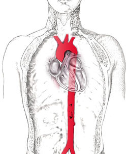
| scheme of the aorta |
| Latin |
aorta |
The aorta (pronounced /eɪˈɔrtə/; from Greek ἀορτή - aortē, from ἀείρω - aeirō "I lift, raise")[1] is the largest artery in the body, originating from the left ventricle of the heart and extends down to the abdomen, where it branches off into two smaller arteries (the common iliacs). The aorta distributes oxygenated blood to all parts of the body through the systemic circulation.[2]
The course of the aorta
The aorta is usually divided into five segments/sections:[3][4]
- Ascending aorta—the section between the heart and the arch of aorta
- Arch of aorta—the peak part that looks somewhat like an inverted "U"
- Descending aorta—the section from the arch of aorta to the point where it divides into the common iliac arteries
- Thoracic aorta—the half of the descending aorta above the diaphragm
- Abdominal aorta—the half of the descending aorta below the diaphragm
In other animals
All amniotes have a broadly similar arrangement to that of humans, albeit with a number of individual variations. In fish, however, there are two separate vessels referred to as aortas. The ventral aorta carries de-oxygenated blood from the heart to the gills; part of this vessel forms the ascending aorta in tetrapods (the remainder forms the pulmonary artery). A second, dorsal aorta carries oxygenated blood from the gills to the rest of the body, and is homologous with the descending aorta of tetrapods. The two aortas are connected by a number of vessels, one passing through each of the gills. Amphibians also retain the fifth connecting vessel, so that the aorta has two parallel arches.[5]
Embryological development
In mammalian and avian embryological development, the pharyngeal arch (aortic arches) arteries contribute to the normal pattern of the great arteries. The fourth aortic arch vessel survives in vertebrates as the arch of the aorta, the third aortic arch vessel persists as the brachiocephalic artery or the root of the internal carotid, and the six arch contributes to the pulmonary arteries. The smooth muscle of the great arteries and the population of cells that form the aorticopulmonary septum that separates the aorta and pulmonary artery is derived from cardiac neural crest. This contribution of the neural crest to the great artery smooth muscle is unusual as most smooth muscle is derived from mesoderm. In fact the smooth muscle within the abdominal aorta is derived from mesoderm, and the coronary arteries, which arise just above the semilunar valves, possess smooth muscle of mesodermal orgin. A failure of the aorticopulmonary septum to divide the great vessels results in persistent truncus arteriosus.
Features
The aorta is an elastic artery, and as such is quite distensible. The blood pressure is highest (normal blood pressure is a reflection of aortic blood pressure) and most pulsatile in the aorta as blood pressure decreases and becomes more smooth (and less pulsatile) as you travel from aorta to arteries to arterioles to capillaries where metabolic exchange occurs. The aorta consists of a heterogeneous mixture of smooth muscle, nerves, intimal cells, endothelial cells, fibroblast-like cells, and a complex extracellular matrix. The vascular wall consists of several layers known as the tunica adventitia, tunica media, and tunica intima. The thickness of the aorta encourages an extensive network of tiny blood vessels called vaso vasorum which feed the layers of the aorta. The aortic arch contains baroreceptors and chemoreceptors which relay information concerning blood pressure and blood pH and carbon dioxide levels to the medulla oblongata of the brain. This information is processed by the brain and the autonomic nervous system mediates the homeostatic responses.
Within the tunica media, smooth muscle and the extracellular matrix are quantitatively the largest components of the aortic vascular wall. The fundamental unit of the aorta is the elastic lamella, which consists of smooth muscle and elastic matrix. The medial layer of the aorta consist of concentric musculoelastic layers (the elastic lamella) in mammals. Functionally the smooth muscle component doesn't dramatically alter the diameter of the aorta but serves to increase the stiffness and viscoelasticity of the aortic wall when activated. The elastic matrix dominates the biomechanical properties of the aorta. The elastic matrix forms lamella consisting of elastic fibers, collagens(predominately type III), proteoglycans, and glycoaminoglycans. When the left ventricle contracts to force blood into the aorta, the aorta expands. This stretching gives the potential energy that will help maintain blood pressure during diastole, as during this time the aorta contracts passively. This Windkessel effect of the great elastic arteries has important biomechanical implications. The elastic recoil helps conserve the energy from the pumping heart and smooth out the pulsatile nature created by the heart. Aortic pressure is highest at the aorta and becomes less pulsatile and lower pressure as blood vessels divide into arteries, arterioles, and capillaries such that flow is slow and smooth for gases and nutrient exchange.
Blood flow and velocity
Clinically, the pulsatile nature of blood flow creates a pulse wave that is propagated down the arterial tree, and at bifurcations reflected waves rebound to return to semilunar valves and the origin of the aorta. These return waves create the dicrotic notch displayed in the aortic pressure curve during the cardiac cycle as these reflected waves push on the aortic semilunar valve. With age the aorta stiffens such the pulse wave is propagated faster and reflected waves return to the heart faster before the semilunar valve closes and this raises the blood pressure. The stiffness of the aorta is associated with a number of diseases and pathologies, and noninvasive measures of the pulse wave velocity are an independent indicator of hypertension. Measuring the pulse wave velocity (invasively and non-invasively) is a means of determining arterial stiffness. Maximum aortic velocity may be noted as Vmax or less commonly as AoVmax.
Anterior (frontal) view of the opened human heart. White arrows indicate normal blood flow.
Diseases/pathology
- Aneurysm of sinus of Valsalva
- Aortic aneurysm - myotic, bacterial (e.g. syphilis), senile, genetic, associated with valvular heart disease
- Aortic coarctation - pre-ductal, post-ductal
- Transposition of the great vessels, see also dextro-Transposition of the great arteries and levo-Transposition of the great arteries
- Atherosclerosis
- Marfan syndrome
- Ehlers-Danlos syndrome
- Aortic stenosis
- Trauma, such as traumatic aortic rupture, most often thoracic and distal to the left subclavian artery[6] and frequently quickly fatal[7]
References
- ↑ Illustrated Steadman's Dictionary, 24th ed.
- ↑ Maton, Anthea (1995). Human Biology Health. Englewood Cliffs, New Jersey: Prentice Hall. ISBN 0-13-981176-1.
- ↑ Tortora, Gerard J: "Principles of Human W. & Karen A. Koos: Human Anatomy, second edition, page 479. Wm. C. Brown Publishing, 1994. (ISBN 0-697-12252-2)
- ↑ De Graaff, Van: "Human Anatomy, fifth edition", pages 548-549. WCB McGraw-Hill, 1998. (ISBN 0-697-28413-1)
- ↑ Romer, Alfred Sherwood; Parsons, Thomas S. (1977). The Vertebrate Body. Philadelphia, PA: Holt-Saunders International. pp. 419–421. ISBN 0-03-910284-X.
- ↑ Samett EJ. http://www.emedicine.com/radio/topic44.htm Aorta, Trauma. eMedicine.com. Accessed on: April 24, 2007.
- ↑ Tambyraja, A; Scollay, JM; Beard, D; Henry, JM; Murie, JA; Chalmers, RT (2006). "Aortic Trauma in Scotland - A Population Based Study". European Journal of Vascular and Endovascular Surgery 32 (6): 686–689. doi:10.1016/j.ejvs.2006.04.006. PMID 16750920.
External links
|
Circulatory system: Arteries and veins (TA A12.0, GA 6.543/GA 7.641) |
|
| Systemic circulation |
|
|
| Pulmonary circulation |
|
|
| Blood vessels |
Endothelium · Tunica intima · Tunica media · Tunica externa
Vasa vasorum · Vasa nervorum
Rete mirabile · Circulatory anastomosis
|
|
| Arteries |
Nutrient artery
|
|
| Veins |
Vena comitans · Superficial vein · Deep vein · Emissary veins · Venous plexus
|
|
| Lymphatic |
Lymph vessel · Lymph · Lymph capillary
|
|
|
|
anat(a:h,u,t,a,l,v:h,u,t,a,l)/phys/devp/cell/
|
|
proc, drug(C2s/,C3,C4,C5,,C8,C9)
|
|
|
|
|
List of arteries of torso · chest (TA A12.2.01-04,11, GA 6.598) |
|
| Pulmonary |
Right pulmonary artery · Left pulmonary artery (Ligamentum arteriosum)
|
|
| Coronary |
Right coronary: SA nodal · AV nodal · Atrial · Right marginal · Posterior interventricular
Left coronary: Anterior interventricular · Left circumflex (Left marginal)
|
|
Ascending aorta/
aortic arch |
|
Brachiocephalic
|
|
|
|
|
|
|
|
Left subclavian
|
Internal thoracic: Anterior intercostal · Thymic · Pericardiacophrenic · Perforating branches · terminal (Musculophrenic, superior epigastric)
Costocervical trunk: Highest intercostal (Posterior intercostal 1-2) · Deep cervical
|
|
|
Other
|
Aortic body
|
|
|
Descending/
thoracic aorta |
visceral: Bronchial · Esophageal · Mediastinal
parietal: Posterior intercostal 3·11 · Subcostal · Superior phrenic
|
|
|
|
anat(a:h,u,t,a,l,v:h,u,t,a,l)/phys/devp/cell/
|
|
proc, drug(C2s/,C3,C4,C5,,C8,C9)
|
|
|
|
|
List of arteries of torso – abdomen (TA A12.2.12-15, GA 6.598) |
|
| AA |
|
Parietal
|
inferior phrenic (superior suprarenal) · lumbar · median sacral · Coccygeal glomus
|
|
|
Anterior
|
|
celiac
|
|
left gastric
|
esophageal branches
|
|
|
common hepatic
|
proper hepatic (cystic) · right gastric · gastroduodenal (right gastro-omental, superior pancreaticoduodenal, supraduodenal)
|
|
|
splenic
|
pancreatic branches (greater, dorsal) · short gastric · left gastro-omental
|
|
|
|
SMA
|
inferior pancreaticoduodenal · intestinal (jejunal, ileal, arcades, vasa recta) · ileocolic (colic, anterior cecal, posterior cecal, ileal branch, appendicular) · right colic · middle colic
|
|
|
IMA
|
left colic · sigmoid · superior rectal · marginal
|
|
|
|
Posterior
|
|
visceral
|
middle suprarenal · renal (inferior suprarenal, ureteral) · gonadal (testicular ♂ / ovarian ♀)
|
|
|
terminal/
common iliac
|
|
IIA
|
|
Anterior
|
|
umbilical
|
superior vesical · to ductus deferens ♂ · medial umbilical ligament
|
|
|
obturator
|
anterior branch · posterior branch
|
|
|
middle rectal
|
vaginal branch ♀
|
|
|
uterine ♀
|
arcuate · vaginal of uterine · ovarian of uterine · tubal of uterine · spiral
|
|
|
V/IV
|
vaginal ♀ · inferior vesical ♂
|
|
|
inferior gluteal
|
accompanying of ischiadic nerve · crucial anastomosis
|
|
|
internal pudendal
|
inferior rectal · perineal(posterior scrotal ♂/labial ♀) · bulb of penis ♂/vestibule ♀ · urethral · deep artery of the penis ♂ (helicine)/clitoris ♀ · dorsal of the penis ♂/clitoris ♀
|
|
|
|
Posterior
|
iliolumbar (lumbar, iliac) · lateral sacral · superior gluteal
|
|
|
|
EIA
|
see arteries of lower limbs
|
|
|
|
|
|
|
anat(a:h,u,t,a,l,v:h,u,t,a,l)/phys/devp/cell/
|
|
proc, drug(C2s/,C3,C4,C5,,C8,C9)
|
|
|
|