Fibula
| Bone: Fibula |
|
|
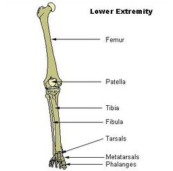
| Bones of lower extremity. |
|
|
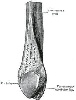
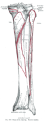
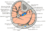
| Figure 1 : Lower extremity of right fibula. Medial aspect. |
| Gray's |
subject #62 260 |
| MeSH |
Fibula |
The fibula or calf bone is a bone located on the lateral side of the tibia, with which it is connected above and below. It is the smaller of the two bones, and, in proportion to its length, the most slender of all the long bones. Its upper extremity is small, placed toward the back of the head of the tibia, below the level of the knee-joint, and excluded from the formation of this joint. Its lower extremity inclines a little forward, so as to be on a plane anterior to that of the upper end; it projects below the tibia, and forms the lateral part of the ankle-joint.
Components
The bone has the following components:
- Body of fibula
- Lateral malleolus
- Interosseous membrane connecting the fibula to the tibia, forming a syndesmoses joint
- The superior tibiofibular articulation is an arthrodial joint between the lateral condyle of the tibia and the head of the fibula.
- The inferior tibiofibular articulation (tibiofibular syndesmosis) is formed by the rough, convex surface of the medial side of the lower end of the fibula, and a rough concave surface on the lateral side of the tibia.
Blood supply
The blood supply is important for planning free tissue transfer because the fibula is commonly used to reconstruct the mandible. The shaft is supplied in its middle third by a large nutrient vessel from the peroneal artery. It is also perfused from its periosteum which receives many small branches from the peroneal artery. The proximal head and the epiphysis are supplied by a branch of the anterior tibial artery. In harvesting the bone the middle third is always taken and the ends preserved (4cm proximally and 6cm distally)
The fibula is ossified from three centers, one for the shaft, and one for either end. Ossification begins in the body about the eighth week of fetal life, and extends toward the extremities. At birth the ends are cartilaginous.
Ossification commences in the lower end in the second year, and in the upper about the fourth year. The lower epiphysis, the first to ossify, unites with the body about the twentieth year; the upper epiphysis joins about the twenty-fifth year.
In other animals
Because the fibula bears relatively little weight in comparison with the tibia, it is typically narrower in all but the most primitive tetrapods. In many animals, it still articulates with the posterior part of the lower extremity of the femur, but this feature is frequently lost (as it is in humans). In some animals, the reduction of the fibula has proceeded even further than it has in humans, with the loss of the tarsal articulation, and, in extreme cases (such as the horse), partial fusion with the tibia.[1]
Additional images
|
|
|
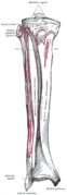
Bones of the right leg. Anterior surface.
|
|
|
|
Bones of the right leg. Posterior surface.
|
|
|
|
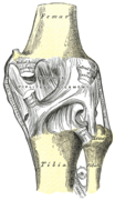
Right knee-joint. Posterior view.
|
|
|
|
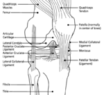
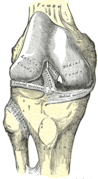
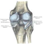
Right knee-joint, from the front, showing interior ligaments.
|
|
|
|
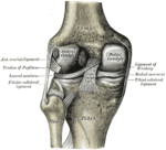
Left knee-joint from behind, showing interior ligaments.
|
|
|
|
Capsule of right knee-joint (distended). Lateral aspect.
|
|
|
|
Capsule of right knee-joint (distended). Posterior aspect.
|
|
|
|
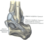
Capsule of left talocrura articulation (distended). Lateral aspect.
|
|
|
|
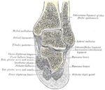
Coronal section through right talocrural and talocalcaneal joints.
|
|
|
|
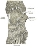
Oblique section of left intertarsal and tarsometatarsal articulations, showing the synovial cavities.
|
|
|
|
Cross-section through middle of leg.
|
|
See also
References
- ↑ Romer, Alfred Sherwood; Parsons, Thomas S. (1977). The Vertebrate Body. Philadelphia, PA: Holt-Saunders International. p. 205. ISBN 0-03-910284-X.
This article was originally based on an entry from a public domain edition of Gray's Anatomy. As such, some of the information contained within it may be outdated.
External links
|
Bones of lower limbs (TA A02.5.04-18, GA 2.242-277) |
|
| Femur |
|
upper extremity
|
head (fovea) · neck · greater trochanter (trochanteric fossa) · lesser trochanter · intertrochanteric line · intertrochanteric crest · quadrate tubercle
|
|
|
body
|
linea aspera · gluteal tuberosity / third trochanter · pectineal line
|
|
|
lower extremity
|
adductor tubercle · patellar surface · epicondyles (lateral, medial) · condyles (lateral, medial) · intercondylar fossa
|
|
|
| Crus |
|
|
|
upper extremity
|
Gerdy's tubercle · condyles (lateral, medial) · intercondylar eminence (lateral/medial intercondylar tubercle) · posterior/anterior intercondylar area
|
|
|
body
|
tuberosity · soleal line
|
|
|
lower extremity
|
medial malleolus · fibular notch
|
|
|
|
Fibula
|
head · body · lateral malleolus
|
|
|
Other
|
|
|
|
| Foot |
|
Tarsus
|
calcaneus (sustentaculum tali, trochlear process) · talus (body, neck, head) · navicular · cuboid · cuneiform (medial, intermediate, lateral)
|
|
|
Metatarsus
|
1st metatarsal · 2nd · 3rd · 4th · 5th
|
|
|
Other
|
phalanges of the foot
|
|
|
|
|
anat(c/f/k/, u, t/p, l)//devp/cell
|
noco/cong/tumr, sysi/, injr
|
|
|
|
|