Humerus
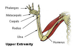
The humerus (ME from Latin humerus, umerus upper arm, shoulder; Gothic ams shoulder, Greek ōmos) is a long bone in the arm or forelimb that runs from the shoulder to the elbow.
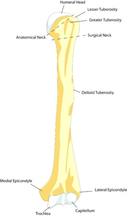
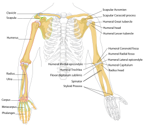
Anatomically, it connects the scapula and the lower arm (consisting of the radius and ulna), and consists of three sections. The upper extremity consists of a rounded head, a narrow neck, and two short processes (tubercles, sometimes called tuberosities.) Its body is cylindrical in its upper portion, and more prismatic below. The lower extremity consists of 2 epicondyles, 2 processes (trochlea & capitulum), and 3 fossae (radial fossa, coronoid fossa, and olecranon fossa)
Muscles attached to the humerus
The deltoid originates on the lateral third of the clavicle, acromion and the spine of the scapula. It is inserted on the deltoid tuberosity of the humerus and has several actions including abduction, extension, and rotation of the shoulder. The supraspinatus also originates on the spine of the scapula. It inserts on the greater tubercle of the humerus, and assists in abduction of the shoulder.
The pectoralis major, teres major, and latissimus dorsi insert at the intertubercular groove of the humerus. They work to adduct and medially, or internally, rotate the humerus.
The infraspinatus and teres minor insert on the greater tubercle, and work to laterally, or externally, rotate the humerus. In contrast, the subscapularis muscle inserts onto the lesser tubercle and works to medially, or internally, rotate the humerus.
The biceps brachii, brachialis, coracobrachialis, and brachioradialis (which attaches distally) act to flex the elbow. (The biceps, however, does not attach to the humerus.) The triceps brachii and anconeus extend the elbow, and attach to the posterior side of the humerus.
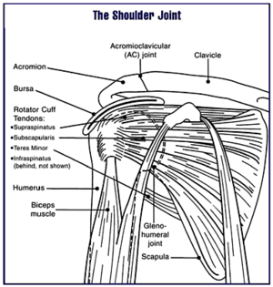
The four muscles of supraspinatus, infraspinatus, teres minor and subscapularis form a musculo-ligamentous girdle called the rotator cuff. This cuff stabilizes the very mobile but inherently unstable glenohumeral joint. The other muscles are used as counterbalances for the actions of lifting/pulling and pressing/pushing.
Articulations
At the shoulder, the head of the humerus articulates with the glenoid fossa of the scapula. More distally, at the elbow, the capitulum of the humerus articulates with the head of the radius, and the trochlea of the humerus articulates with the olecranon process of the ulna.
Nerves
The axillary nerve is located at the proximal end, against the shoulder girdle. The most common type of shoulder dislocation is an anterior or inferior dislocation of the humerus's glenohumeral joint, which has the potential to injure the axillary nerve or the axillary artery. Signs and symptoms of this dislocation include a loss of the normal shoulder contour and a palpable depression under the acromion.
The radial nerve follows the humerus closely. At the midshaft of the humerus, the radial nerve travels from the posterior to the anterior aspect of the bone in the spiral groove. A fracture of the humerus in this region can result in radial nerve injury.
The ulnar nerve at the distal end of the humerus near the elbow is sometimes referred to in popular culture as 'the funny bone'. Striking this nerve can cause a tingling sensation ("funny" feeling), and sometimes a significant amount of pain.
In other animals
Primitive fossil amphibians had little, if any, shaft connecting the upper and lower extremities, making their limbs very short. In most living vertebrates, however, the humerus has a similar form to that of humans. In many reptiles and some primitive mammals, the lower extremity includes a large foramen, or opening, into which nerves and blood vessels pass.[1]
Additional images
|
|
|
Diagram of the human shoulder joint
|
|
|
|
Humerus (right) - anterior view
|
|
|
|
Humerus (right) - posterior view
|
|
|
|
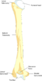
Left humerus. Anterior view.
|
|
|
|
Left humerus. Posterior view.
|
|
|
|
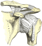
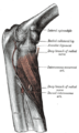
The left shoulder and acromioclavicular joints, and the proper ligaments of the scapula.
|
|
|
|
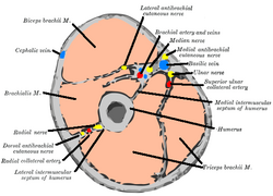
Cross-section through the middle of upper arm.
|
|
References
- ↑ Romer, Alfred Sherwood; Parsons, Thomas S. (1977). The Vertebrate Body. Philadelphia, PA: Holt-Saunders International. pp. 198–199. ISBN 0-03-910284-X.
This article was originally based on an entry from a public domain edition of Gray's Anatomy. As such, some of the information contained within it may be outdated.
|
Bones of upper limbs (TA A02.4, GA 2.200-230) |
|
Pectoral girdle,
clavicle |
conoid tubercle · trapezoid line · costal tuberosity · subclavian groove
|
|
| Scapula |
fossae (subscapular, supraspinatous, infraspinatous) · suprascapular notch · glenoid cavity
tubercles (infraglenoid, supraglenoid) · spine of scapula · acromion · coracoid process
borders (superior, lateral/axillary, medial/vertebral) · angles (superior, inferior, lateral)
|
|
| Humerus |
upper extremity: necks (anatomical, surgical) · tubercles (greater, lesser) · intertubercular sulcus
body: radial sulcus · deltoid tuberosity
lower extremity: capitulum · trochlea · epicondyles (lateral, medial) · supracondylar ridges (lateral, medial) · fossae (radial, coronoid, olecranon)
|
|
| Forearm |
radius: upper extremity (head, tuberosity) · body · lower extremity (ulnar notch, styloid process)
ulna: upper extremity (tuberosity, olecranon, coronoid process, radial notch, trochlear notch) · body · lower extremity (head, styloid process)
|
|
| Hand |
carpus: scaphoid · lunate · triquetral · pisiform · trapezium · trapezoid · capitate · hamate (hamulus)
metacarpus: 1st metacarpal · 2nd · 3rd · 4th · 5th
phalanges of the hand: proximal · intermediate · distal
|
|
|
|
anat(c/f/k/, u, t/p, l)//devp/cell
|
noco/cong/tumr, sysi/, injr
|
|
|
|
|