Pons
| Brain: Pons | ||
|---|---|---|
 |
||
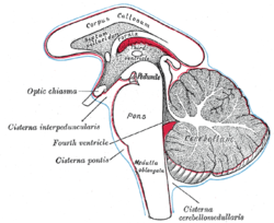
| Diagram showing the positions of the three principal subarachnoid cisternæ (pons visible at center) | ||
 |
||
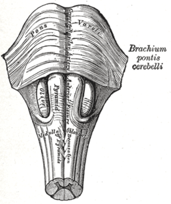
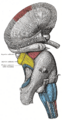
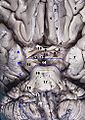
| Anteroinferior view of the medulla oblongata and pons | ||
| Gray's | subject #187 785 | |
| Part of | Brain stem | |
| Artery | pontine arteries | |
| Vein | transverse and lateral pontine veins | |
| NeuroNames | hier-538 | |
| MeSH | Pons | |
| NeuroLex ID | birnlex_733 | |
The pons (Latin for "bridge"), sometimes pons Varolii (after Costanzo Varolio, a 16th-century Italian anatomist and surgeon) is a structure located on the brain stem. It is superior to (up from) the medulla oblongata, inferior to (down from) the midbrain, and rostral to (in front of) the cerebellum. In humans and other bipeds this means it is above the medulla, below the midbrain, and anterior to the cerebellum. Its white matter includes tracts that conduct signals from the cerebrum down to the cerebellum and medulla, and tracts that carry the sensory signals up into the thalamus.[1]
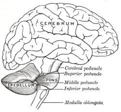
The pons measures about 2.5 cm in length. Most of it appears as a broad anterior bulge rostral to the medulla. Posteriorly, it consists mainly of two pairs of thick stalks called cerebellar peduncles. They connect the cerebellum to the pons and midbrain. [2]
The pons contains nuclei that relay signals from the cerebrum to the cerebellum, along with nuclei that deal primarily with sleep, respiration, swallowing, bladder control, hearing, equilibrium, taste, eye movement, facial expressions, facial sensation, and posture.[3]
Within the pons is the pneumotaxic center, a nucleus in the pons that regulates the change from inspiration to expiration.[4]
Contents |
Embryonic development
During embryonic development the embryonic metencephalon develops into two structures: the pons and the cerebellum.[5]
Cranial nerve nuclei
A number of cranial nerve nuclei are present in the pons:
- mid-pons: The chief or pontine nucleus of the trigeminal nerve sensory nucleus (V)
- mid-pons: the motor nucleus for the trigeminal nerve (V)
- lower down in the pons: abducens nucleus (VI)
- lower down in the pons: facial nerve nucleus (VII)
- lower down in the pons: vestibulocochlear nuclei (vestibular nuclei and cochlear nuclei) (VIII)
The functions of these four nerves include sensory roles in hearing, equilibrium, and taste, and in facial sensations such as touch and pain; as well as motor roles in eye movement, facial expressions, chewing, swallowing, urination, and the secretion of saliva and tears. [6]
Related diseases
- Central pontine myelinosis, a demyelination disease that causes difficulty with sense of balance, walking, sense of touch, swallowing and speaking. In a clinical setting it is often associated with transplant. Undiagnosed it can lead to death or locked-in syndrome.
Additional images
 Location and topography of Pons(Animation) |
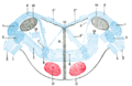
 Scheme showing the connections of the several parts of the brain. |
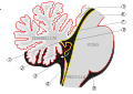
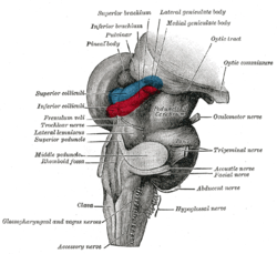
 Superficial dissection of brain-stem. Lateral view. |
 Superficial dissection of brain-stem. Ventral view. |
 Axial section of the pons, at its upper part. |
 Dissection showing the projection fibers of the cerebellum. |
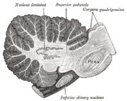 Sagittal section through right cerebellar hemisphere. The right olive has also been cut sagitally. |
 Scheme of roof of fourth ventricle. The arrow is in the foramen of Majendie. |
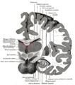
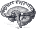
 Mesal aspect of a brain sectioned in the median sagittal plane. |
 Coronal section of brain immediately in front of pons. |
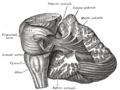
 Hind- and mid-brains; postero-lateral view. |
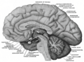
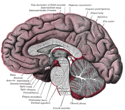 Median sagittal section of brain. |
 Dissection showing the course of the cerebrospinal fibers. |
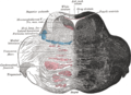
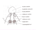
 Terminal nuclei of the cochlear nerve, with their upper connections. |
 Terminal nuclei of the vestibular nerve, with their upper connections. |
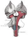
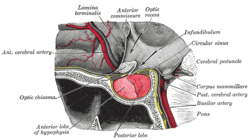 The hypophysis cerebri in position. Shown in sagittal section. |
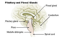 Pituitary and pineal glands |
 Axial section of the Brainstem (Pons) at the level of the Facial Colliculus |
 Human cerebrum lateral view |
_section_description.JPG) Human brain frontal (coronal) section |
 Human brainstem anterior view |
References
Saladin Kenneth S.(2007) Anatomy & physiology the unity of form and function. Dubuque, IA: McGraw-Hill
External links
- Diagram at UCC
- BrainMaps at UCDavis Pons
|
||||||||||||||||||||||||||||
|
||||||||||||||||||||||||||||||||||||||||||