Scabies
| Scabies | |
|---|---|
| Classification and external resources | |
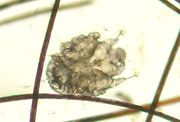
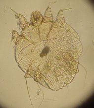 A photomicrograph of an itch mite (Sarcoptes scabiei). |
|
| ICD-10 | B86. |
| ICD-9 | 133.0 |
| DiseasesDB | 11841 |
| MedlinePlus | 000830 |
| eMedicine | derm/382 emerg/517 ped/2047 |
| MeSH | D012532 |
Scabies, also known as sarcoptic mange and colloquially known as the itch, is a contagious ectoparasitic skin infection characterized by superficial burrows and intense pruritus (itching). It is caused by the mite Sarcoptes scabiei. The word scabies itself is derived from the Latin word (scabere) for "scratch". More severe forms of scabies include Norwegian scabies and crusted scabies.
Contents |
Signs and symptoms
The characteristic symptoms of scabies infection include superficial burrows, intense pruritus (itching), a generalized rash and secondary infection. Acropustulosis, or blisters and pustules on the palms and soles of the feet, are characteristic symptoms of scabies in infants.[1]
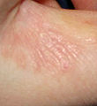
S-shaped tracks in the skin are often accompanied by small, insect-type bites called nodules that may look like pimples.[1] These burrows and nodules are often located in the crevices of the body, such as the webs of fingers, toes, feet, buttocks, elbows, waist area, genital area and axilla, and under the breasts in women.[1]
The intense itching and rash characteristic of scabies infection is caused by an allergic reaction of the body to the burrowed microscopic scabies mites. The rash can be found over much of the body, especially in immunocompromised people (HIV positive or elderly); the associated itching is often most prevalent at night.[2]
Secondary infection of impetigo, a Streptococci or Staphylococci bacterial skin infection, may occur after scratching. Cellulitis may also occur, resulting in localized swelling, redness and fever (DermNet).
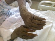
In immuno-compromised, malnourished, elderly or institutionalized individuals, infestation can cause a more severe form of scabies known as crusted scabies or Norwegian scabies. This syndrome is characterized by a scaly rash, slight itching and thickened crusts of skin containing thousands of mites.[2] Norwegian scabies is the form of scabies that is hardest to treat.
In individuals never before exposed to scabies, the onset of clinical signs and symptoms is 4–6 weeks after infestation. Some people may not realize that they have it for years; in previously exposed individuals, onset can be as soon as 2–4 days after infestation.
Compromised immune systems
People with compromised immune systems, such as people with HIV or cancer or transplant patients on immunosuppressive drugs, may be susceptible to Crusted (Norwegian) scabies. In this case the scabies go unregulated by cytotoxic T cells and spread over the whole body, except the face. These cases require additional treatment options for resolution. Ivermectin is a single oral treatment of choice in these patients combined with any other topical treatment.
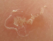
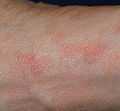
Gallery
 Scabies of the foot |
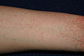 Scabies of the arm |
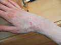
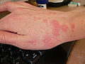
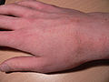
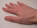
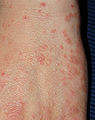 Scabies of the hand |
 Scabies of the finger |
Evolution of infection
 Day 4 |
 Day 8 (treatment begins) |
 Day 12 (under treatment) |
 Healed |
Cause
Scabies is highly contagious and can be spread by scratching, picking up the mites under the fingernails and simply touching another person's skin. They can also be spread onto other objects like keyboards, toilets, clothing, towels, bedding, furniture, and anything else onto which the mite may be rubbed off, especially if a person is heavily infested. The parasite can survive up to 14 days away from a host, but often do not survive longer than two or three days away from human skin.[3] Scabies is caused by the mite Sarcoptes scabiei, variety hominis, as shown by the Italian biologist Diacinto Cestoni in the 18th century. It produces intense, itchy skin rashes when the impregnated female tunnels into the stratum corneum of the skin and deposits eggs in the burrow. The larvae, which hatch in 3–10 days, move about on the skin, molt into a "nymphal" stage, and then mature into adult mites. The adult mites live 3–4 weeks in the host's skin.
The action of the mites moving within the skin and on the skin itself produces an intense itch which may resemble an allergic reaction in appearance. The presence of the eggs does not, in fact, produce more itching; this conjecture is only myth. It is rather the feces of the mites which cause the allergic reaction.
Scabies can be transmitted readily throughout an entire household by skin-to-skin contact with an infected person (e.g. bed partners, schoolmates, daycare). It can be spread by clothing, bedding or towels. Washing clothing in very hot water and dry on high heat will help prevent the transmission. Alternatively, permethrin sprays can be used for items that cannot be laundered.
The symptoms of itching and rash are caused by an allergic reaction that the human body develops over time to the mites and their by-products under the skin. As such, there is usually a 2-6 week incubation period between infestation and presentation of symptoms. However, in individuals with prior exposure to scabies, the incubation period is much shorter: as little as 1–4 days.[4]
There are usually relatively few mites on a normal, healthy person (who is infested with scabies) — about 11 females in burrows. Scabies are microscopic although sometimes they are visible as a pinpoint of white. The females burrow into the skin and lay eggs there. Males roam on top of the skin but can also occasionally burrow.
Diagnosis
Signs and symptoms of early scabies infestation mirror other skin diseases, including dermatitis, syphilis, allergic reactions and other ectoparasites such as lice and fleas.[5]
Generally diagnosis is made by finding burrows. This may be difficult because they are scarce and because they are obscured by scratch marks. If burrows are not found in the primary areas known to be affected, the entire skin surface of the body should be examined.
The suspicious area can be rubbed with ink from a fountain pen or a topical tetracycline solution which will glow under a special light. The surface is then wiped off with an alcohol pad; if the person is infected with scabies, the characteristic zigzag or "S" pattern of the burrow across the skin will appear.
When a suspected burrow is found, diagnosis may be confirmed by microscopy of surface scrapings taken with a scalpel or curette. These are then placed on a slide in glycerol or mineral oil and contained with a coverslip. Avoiding potassium hydroxide is necessary because it may dissolve fecal pellets. Positive diagnosis is made when the mite, ova or fecal pellets are found. Although this sounds simple in practice, actual detection of scabies sites is very difficult. It often requires the scraping of dozens of suspicious lesions down to the superficial dermis. This will result in minor bleeding in spots. Even a negative (unsuccessful) scraping will not completely rule out scabies. Sometimes the best diagnosis is by the history, physical findings and response to effective topical treatment. The diagnosis of Crusted Scabies is not as elusive and a scraping under the fingernail is often diagnostic. Scabies manifests as papules, pustules, burrows, nodules, and occasionally urticarial papules and plaques. Most of the patients with scabies experience severe pruritus.[6]
Management
Medications
- Sulphur has been used since around 25 AD to treat Scabies. Bar soap is commercially available with sulfur in the ranges of 1%-10% to kill scabies. 6% or above is recommended. Wash the whole body at least once daily for 4 consecutive days. And although supposedly minimally effective it is still recommended that all clothing, bed linen and blankets be washed in hot water and dried in high heat and then treated with Permethrin.
Topical drugs
- Permethrin 5% is topical medication of choice.[7] Toxicity may resemble allergic reactions. It is usually applied to the skin before bedtime and left on for about 8 to 14 hours, then showered off in the morning. Package directions or doctor's instructions should be followed, but one application is normally sufficient to cure an infection.[8]
- Eurax (USP Crotamiton) This is a cure and also helps to relieve itch (pruritis)[9]
- Malathion: Applied for 24 hours; effective in killing both adults and eggs.
- Lindane lotion is approved in the U.S. for use as a second-line treatment where first-line medications like permethrin have either failed, are not well tolerated or otherwise contraindicated.[10] It is illegal in 17 other countries, and 33 more countries have restricted its use.[11][12] Though rare, serious side effects have resulted from product misuse.[11][13] The FDA has confirmed 3 deaths that all involved use of lindane not in accordance with the label, including excessive topical applications and oral ingestions.[14]
- There is some evidence that a 10% sulfur ointment in petroleum jelly applied topically is effective. It is cheap and readily available without a prescription.[15][16] It also has the advantage of being well-tolerated in pregnant women and infants under two months of age.
- Neem oil is deemed very effective in the treatment of scabies although only preliminary scientific proof exists which has yet to be corroborated. It is recommended for those who are sensitive to permethrin, a known insecticide, which might cause irritation. The scabies mite, furthermore, has yet to become resistant to neem, so in persistent cases neem has been shown to be very effective.[17]
- Tea tree oil at 5% was only partially effective and does not seem to be a viable solution for treatment. In one study, it was more effective than commercial medications against the scabies mite in an in vitro situation.[18]
Oral
A single dose of Ivermectin has been reported to reduce the load of scabies but another dose is required after 2 weeks for full eradication. In 1999 a small scale test comparing topically applied Lindane to orally administered Ivermectin found no statistically significant differences between the two treatments.[19] As Ivermectin is easily administered (not requiring a rub-down of the whole body like lindane or permethrin twice per treatment), compliance is much higher among self-administering patients. Ivermectin is used in eradication programs of many parasites of both human and animal. Side effects may include mild abdominal pain, nausea, vomiting, myalgia and/or arthralgia, but they all usually diminish even while treatment is still underway. The product is considered safe for use in children over five years of age.[20]
Public health and prevention strategies
There is no vaccine available for scabies, nor are there any proven causative risk factors. Therefore, most strategies focus on preventing re-infection. All family and close contacts should be treated at the same time, even if asymptomatic. Cleaning of environment should occur simultaneously, as there is a risk of reinfection. Therefore it is recommended to wash and hot iron all material (such as clothes, bedding and towels) that has been in proximity to a scabies infestation.
Cleaning of the environment should include:
- Treatment of furniture and bedding.
- Vacuuming floors, carpets and rugs.
- Disinfecting floor and bathroom surfaces by mopping.
- Cleaning the shower/bath tub after each use.
- Daily washing of recently worn clothes, towels and bedding in hot water and then drying in high heat and/or steam ironing.
Itchiness during treatment
Options to combat itchiness include antihistamines such as chlorpheniramine. Prescription: Hydroxyzine (Atarax).
Epidemiology
Scabies is impressively egalitarian in its epidemiology. Mites are distributed around the world, affecting all ages, races and socioeconomic classes in all different climates.[2] It is more often seen, however, in crowded and unhygienic living conditions.[21] Globally there is an estimated incidence of 300 million cases of scabies per year, 1 million of which occur in the United States.[4]
History
Scabies is an ancient disease. Based on archeological evidence from Egypt and the Middle East, scabies is estimated to date back over 2,500 years.[4] The first recorded reference to scabies is believed to be from the Bible (Leviticus, the third book of Moses) ca. 1200 BC. Later, the ancient Greek philosopher Aristotle reported on “lice” that would “escape from little pimples if they are pricked” in the fourth century BC;[22] scholars believe this was actually a reference to scabies.
Nevertheless it was the Roman physician Celsus who is credited with designating the term “scabies” to the disease and describing its characteristic features.[22] The parasitic etiology of scabies was later documented by the Italian physician Giovanni Cosimo Bonomo (1663-1699 AD) in his famous 1687 letter, “Observations concerning the fleshworms of the human body.”[22] With this (disputed) discovery, scabies became one of the first diseases with a known cause.[4]
In 1844 Ferdinand von Hebra discovered the cause of scabies. (http://www.whonamedit.com/doctor.cfm/708.html)
Animals

Many domestic animals have their own species of Sarcoptes mites. Though all can transiently affect humans,[23][24] the mites that cause scabies in animals cannot reproduce on the human body. Humans are especially susceptible to small dogs carrying the mites. Recent outbreaks have started to reach epidemic proportions.[25] The most frequently diagnosed form is sarcoptic mange in dogs. In dogs and other animals, scabies produces severe itching and secondary skin infections. Affected animals often lose weight and become unthrifty. Sarcoptes is a genus of skin parasites and part of the larger family of mites collectively known as “scab mites”. They are also related to the scab mite Psoroptes that infests the skin of domestic animals. Sarcoptic mange affects domestic animals and similar infestations in domestic fowls causes the disease known as “scabies leg”. The effects of Sarcoptes scabiei are the most well known, causing “scabies”, or “the itch”. The adult female mite, having been fertilised, burrows into the skin, usually the hands or wrists, and then lays her eggs. Other parts of the body may also be affected.
Scabies has been observed on non-domestic animals as well. Gorillas, for instance, are known to be susceptible to infection via contact with items used by humans.[26]
Feral animals
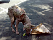
Domestic animals that have gone feral and have no veterinary care are frequently afflicted with scabies and a host of other ailments.[27]
See also
- Crab louse
- Head louse
- List of parasites (human)
- List of cutaneous conditions
References
- ↑ 1.0 1.1 1.2 "Scabies". DermNet NZ. New Zealand Dermatological Society Incorporated. http://www.dermnetnz.org/arthropods/pdf/scabies-dermnetnz.pdf.
- ↑ 2.0 2.1 2.2 "DPDx - Scabies". Laboratory Identification of Parasites of Public Health Concern. CDC. http://www.dpd.cdc.gov/dpdx/HTML/Scabies.htm.
- ↑ http://www.cdc.gov/scabies
- ↑ 4.0 4.1 4.2 4.3 Markell, Edward K.; John, David C.; Petri, William H. (2006). Markell and Voge's medical parasitology (9th ed.). St. Louis, Mo: Elsevier Saunders. ISBN 0-7216-4793-6.
- ↑ Arlian LG (1989). "Biology, host relations, and epidemiology of Sarcoptes scabiei". Annu. Rev. Entomol. 34: 139–61. doi:10.1146/annurev.en.34.010189.001035. PMID 2494934. http://arjournals.annualreviews.org/doi/abs/10.1146/annurev.en.34.010189.001035?url_ver=Z39.88-2003&rfr_id=ori:rid:crossref.org&rfr_dat=cr_pub%3dncbi.nlm.nih.gov.
- ↑ Scheinfeld N. Controlling scabies in institutional settings: a review of medications, treatment models, and implementation. Am J Clin Dermatol. 2004;5:31-7. "PMID 14979741"
- ↑ Scheinfeld NS (2004). "Controlling scabies in institutional settings: a review of medications, treatment models, and implementation". Amer J Clin Dermatol 5 (1): 31–7. doi:10.2165/00128071-200405010-00005. PMID 14979741.
- ↑ [1]
- ↑ http://www.nlm.nih.gov/medlineplus/druginfo/uspdi/202170.html
- ↑ FDA Public Health Advisory: Safety of Topical Lindane Products for the Treatment of Scabies and Lice
- ↑ 11.0 11.1 http://www.fda.gov/cder/foi/label/2003/006309lotionlbl.pdf.
- ↑ Commission for Environmental Cooperation. North American Regional Action Plan (NARAP) on lindane and other hexachlorocyclohexane (HCH) isomers. November 30, 2006.
- ↑ "Lindane Post Marketing Safety Review" (PDF). U.S. Food and Drug Administration (FDA). 2003. http://www.fda.gov/cder/drug/infopage/lindane/lindaneaeredacted.pdf.
- ↑ http://www.fda.gov/Drugs/DrugSafety/PublicHealthAdvisories/ucm052201.htm
- ↑ Lin AN, Reimer RJ, Carter DM (1988). "Sulfur revisited". J Am Acad Dermatol 18: 553–58. doi:10.1016/S0190-9622(88)70079-1.
- ↑ Pruksachatkunakorn C, Damrongsak M, Sinthupuan S (2002). "Sulfur for Scabies Outbreaks in Orphanages". Pediatric Dermatology 19 (5): 448–53. doi:10.1046/j.1525-1470.2002.00205.x. PMID 12383106. http://www3.interscience.wiley.com/journal/118919953/abstract. Retrieved 2008-08-01.
- ↑ Heinrich M, et al. (2005). "Plants as Medicines". In Prance G, Nesbitt M. The Cultural History of Plants. London: Routledge. pp. 228. ISBN 0415927463.
- ↑ Walton SF, McKinnon M, Pizzutto S, Dougall A, Williams E, Currie BJ (May 2004). "Acaricidal activity of Melaleuca alternifolia (tea tree) oil: in vitro sensitivity of sarcoptes scabiei var hominis to terpinen-4-ol". Arch Dermatol 140 (5): 563–6. doi:10.1001/archderm.140.5.563. PMID 15148100.
- ↑ Apgar B (January 15, 2000). "Efficacy and Safety of Therapy for Human Scabies Infestation". American Family Physician. http://www.aafp.org/afp/20000115/tips/15.html.
Chouela EN, Abeldaño AM, Pellerano G, et al. (June 1999). "Equivalent therapeutic efficacy and safety of ivermectin and lindane in the treatment of human scabies". Arch Dermatol 135 (6): 651–5. doi:10.1001/archderm.135.6.651. PMID 10376691. http://archderm.ama-assn.org/cgi/pmidlookup?view=long&pmid=10376691.
Strong M, Johnstone PW (2007). "Interventions for treating scabies". Cochrane Database Syst Rev (3): CD000320. doi:10.1002/14651858.CD000320.pub2. PMID 17636630. - ↑ Borsboom GJ, Boatin BA, Nagelkerke NJ, et al. (March 2003). "Impact of ivermectin on onchocerciasis transmission: assessing the empirical evidence that repeated ivermectin mass treatments may lead to elimination/eradication in West-Africa". Filaria J 2 (1): 8. doi:10.1186/1475-2883-2-8. PMID 12769825. PMC 156613. http://www.filariajournal.com/content/2/1/8.
- ↑ Green MS (1989). "Epidemiology of scabies". Epidemiol Rev 11: 126–50. PMID 2509232. http://epirev.oxfordjournals.org/cgi/reprint/11/1/126.
- ↑ 22.0 22.1 22.2 Roncalli RA (July 1987). "The history of scabies in veterinary and human medicine from biblical to modern times". Vet. Parasitol. 25 (2): 193–8. doi:10.1016/0304-4017(87)90104-X. PMID 3307123. http://linkinghub.elsevier.com/retrieve/pii/0304-4017(87)90104-X.
- ↑ Chakrabarti A (1985). "Some epidemiological aspects of animal scabies in human population". Int J Zoonoses 12 (1): 39–52. PMID 4055268.
- ↑ Ulmer A, Schanz S, Röcken M, Fierlbeck G (2007). "A papulovesicular rash in a farmer and his wife". Clin Infect Dis 45 (3): 395–96. doi:10.1086/519434. PMID 17599314.
- ↑ Scabies at the US Centers for Disease Control and Prevention
- ↑ http://www.pbs.org/wgbh/pages/frontline/story/2009/11/doomsday-thinking.html
- ↑ "Bali Animal Welfare Association". http://www.bawabali.com/. Retrieved 2009-07-28.
External links
- American Academy of Dermatology pamphlet on Scabies
- Scabies FAQ from the National Pediculosis Association
|
|||||||||||||||||||||||||||||||||||||||||||||||||||||||||||||||||||||||||||
|
||||||||||||||||||||||||||||||||||||||