Uterus
| Uterus | |
|---|---|
 |
|
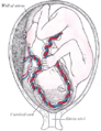
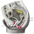
| 1. Round ligament 2. Uterus 3. Uterine cavity 4. Intestinal surface of Uterus 5. Versical surface(toward bladder) 6. Fundus of uterus 7. Body of uterus 8. Palmate folds of cervical canal 9. Cervical canal 10. Posterior lip 11. Cervical os (external) 12. Isthmus of uterus 13. Supravaginal portion of cervix 14. Vaginal portion of cervix 15. Anterior lip 16. Cervix |
|
| Gray's | subject #268 1258 |
| Artery | ovarian artery, uterine artery, helicine branches of uterine artery |
| Vein | uterine veins |
| Lymph | body and cervix to internal iliac lymph nodes, fundus to para-aortic lymph nodes |
| Precursor | Müllerian duct |
| MeSH | Uterus |
The uterus (from Latin "uterus" (womb, belly), plural or uteri) or womb is a major female hormone-responsive reproductive sex organ of most mammals including humans. One end, the cervix, opens into the vagina, while the other is connected to one or both fallopian tubes, depending on the species. It is within the uterus that the fetus develops during gestation, usually developing completely in placental mammals such as humans and partially in marsupials such as kangaroos and opossums. Two uteruses usually form initially in a female fetus, and in placental mammals they may partially or completely fuse into a single uterus depending on the species. In many species with two uteruses, only one is functional. Humans and other higher primates such as chimpanzees, along with horses, usually have a single completely fused uterus, although in some individuals the uteruses may not have completely fused. The term uterus is used consistently within the medical and related professions, while the Germanic derived term womb is also common in everyday usage in the English language.
Most animals that lay eggs, such as birds and reptiles, have an oviduct instead of a uterus. In monotremes, mammals which lay eggs and include the platypus, either the term uterus or oviduct is used to describe the same organ, but the egg does not develop a placenta within the mother and thus does not receive further nourishment after formation and fertilization. Marsupials have two uteruses, each of which connect to a lateral vagina and which both use a third, middle "vagina" which functions as the birth canal. Marsupial embryos form a choriovitelline "placenta" (which can be thought of as something between a monotreme egg and a "true" placenta), in which the egg's yolk sac supplies a large part of the embryo's nutrition but also attaches to the uterine wall and takes nutrients from the mother's bloodstream.
Contents |
Function
The uterus provides structural integrity and support to the bladder, bowel, pelvic bones and organs. The uterus helps separate and keep the bladder in its natural position above the pubic bone and the bowel in its natural configuration behind the uterus. The uterus is continuous with the cervix, which is continuous with the vagina, much in the way that the head is continuous with the neck, which is continuous with the shoulders. It is attached to bundles of nerves, and networks of arteries and veins, and broad bands of ligaments such as round ligaments, cardinal ligaments, broad ligaments, and uterosacral ligaments.[1]
The uterus is essential in sexual response by directing blood flow to the pelvis and to the external genitalia, including the ovaries, vagina, labia, and clitoris. The uterus is needed for uterine orgasm to occur.
The reproductive function of the uterus is to accept a fertilized ovum which passes through the utero-tubal junction from the fallopian tube. It then becomes implanted into the endometrium, and derives nourishment from blood vessels which develop exclusively for this purpose. The fertilized ovum becomes an embryo, attaches to a wall of the uterus, creates a placenta, and develops into a fetus (gestates) until childbirth. Due to anatomical barriers such as the pelvis, the uterus is pushed partially into the abdomen due to its expansion during pregnancy. Even during pregnancy the mass of a human uterus amounts to only about a kilogram (2.2 pounds).
Forms in mammals
In mammals, the four main forms in which it is found are:
- Duplex
- There are two wholly separate uteri, with one fallopian tube each. Found in marsupials (such as kangaroos, Tasmanian devils, opossums, etc.), rodents (such as mice, rats, and guinea pigs), and lagomorpha (rabbits and hares).
- Bipartite
- The two uteri are separate for most of their length, but share a single cervix. Found in ruminants (deer, moose, elk etc.), and cats.
- Bicornuate
- The upper parts of the uterus remain separate, but the lower parts are fused into a single structure. Found in dogs, pigs, elephants, whales, dolphins, and prosimian primates among others.
- Simplex
- The entire uterus is fused into a single organ. Found in higher primates (including humans and chimpanzees) and horses. Occasionally, some individual females (including humans) may have a bicornuate uterus, a uterine malformation where the two parts of the uterus fail to fuse completely during fetal development.
In monotremes such as the platypus, the uterus is duplex and rather than nurturing the embryo, secretes the shell around the egg. It is essentially identical with the shell gland of birds and reptiles, with which the uterus is homologous.[2]
Anatomy
The uterus is located inside the pelvis immediately dorsal (and usually somewhat rostral) to the urinary bladder and ventral to the rectum. The human uterus is pear-shaped and about 3 in. (7.6 cm) long. A female's uterus can be divided anatomically into four segments: The fundus, corpus, cervix and the internal os.
Regions
From outside to inside, the path to the uterus is as follows:
- Cervix uteri - "neck of uterus"
- External orifice of the uterus
- Canal of the cervix
- Internal orifice of the uterus
- corpus uteri - "Body of uterus"
- Cavity of the body of the uterus
- Fundus (uterus)
Layers
The layers, from innermost to outermost, are as follows:
- Endometrium
- The lining of the uterine cavity is called the "endometrium". It consists of the functional endometrium and the basal endometrium from which the former arises. Damage to the basal endometrium results in adhesion formation and/or fibrosis (Asherman's syndrome). In all placental mammals, including humans, the endometrium builds a lining periodically which is shed or reabsorbed if no pregnancy occurs. Shedding of the functional endometrial lining is responsible for menstrual bleeding (known colloquially as a "period" in humans with a cycle of about 28 days) throughout the fertile years of a female and for some time beyond. Depending on the species, menstrual cycles may vary from a few days to six months, but can vary widely even in the same individual, often stopping for several cycles before resuming. Marsupials and monotremes do not have menstruation.
- Myometrium
- The uterus mostly consists of smooth muscle, known as "myometrium." The innermost layer of myometrium is known as the junctional zone, which becomes thickened in adenomyosis.
- Perimetrium
- The loose surrounding tissue is called the "perimetrium."
- Peritoneum
- The uterus is surrounded by "peritoneum."
Support
The uterus is primarily supported by the pelvic diaphragm, perineal body and the urogenital diaphragm. Secondarily, it is supported by ligaments and the peritoneum (broad ligament of uterus) [3]
Major ligaments
It is held in place by several peritoneal ligaments, of which the following are the most important (there are two of each):
| Name | From | To |
|---|---|---|
| Uterosacral ligament | Posterior cervix | Sacrum of pelvis |
| Cardinal ligaments | Side of the cervix | Ischial spines |
| Pubocervical ligament [3] | Side of the cervix | Pubic symphysis |
Other named ligaments near the uterus, i.e. the broad ligament, the round ligament, the suspensory ligament of the ovary, the infundibulopelvic ligament, have no role in the support of the uterus.
Position
Under normal circumstances the uterus is both "anteflexed" and "anteverted." The meaning of these terms are described below:
| Distinction | More common | Less common |
|---|---|---|
| Position tipped | "Anteverted": Tipped forward | "Retroverted": Tipped backwards |
| Position of fundus | "Anteflexed": Fundus is pointing forward relative to the cervix | "Retroflexed": Fundus is pointing backwards |
Development
The bilateral Müllerian ducts form during early fetal life. In males, MIF secreted from the testes leads to their regression. In females these ducts give rise to the Fallopian tubes and the uterus. In humans the lower segments of the two ducts fuse to form a single uterus, however, in cases of uterine malformations this development may be disturbed. The different uterine forms in various mammals are due to various degrees of fusion of the two Müllerian ducts.
Sexual response
Stimulation of the uterus can result in an intense orgasm.
Pathology
Some pathological states include:
- Prolapse of the uterus
- Carcinoma of the cervix – malignant neoplasm
- Carcinoma of the uterus – malignant neoplasm
- Fibroids – benign neoplasms
- Adenomyosis – ectopic growth of endometrial tissue within the myometrium
- Pyometra – infection of the uterus, most commonly seen in dogs
- Uterine malformations mainly congenital malformations including Uterine Didelphys, bicornuate uterus and septate uterus. It also includes congenital absence of the uterus Rokitansky syndrome
- Asherman's syndrome, also known as intrauterine adhesions occurs when the basal layer of the endometrium is damaged by instrumention (e.g. D&C) or infection (e.g. endometrial tuberculosis) resulting in endometrial scarring followed by adhesion formation which partially or completely obliterates the uterine cavity.
Additional images
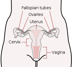 Schematic frontal view of female anatomy |
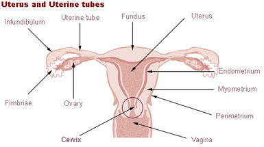 Uterus and uterine tubes. |
 |
 Anatomical model of a human pregnancy |
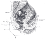
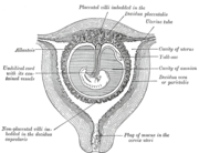 Sectional plan of the gravid uterus in the third and fourth month. |
 Fetus in utero, between fifth and sixth months. |
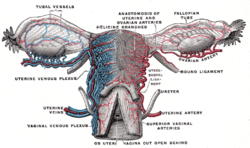 Vessels of the uterus and its appendages, rear view. |
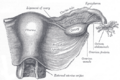 Uterus and right broad ligament, seen from behind. |
 Female pelvis and its contents, seen from above and in front. |
 Sagittal section of the lower part of a female trunk, right segment. |
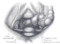
Posterior half of uterus and upper part of vagina. |
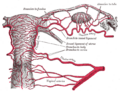 The arteries of the internal organs of generation of the female, seen from behind. |
 Median sagittal section of female pelvis. |
See also
- Hysterectomy
- Menopause
- Uterine glands
References
- ↑ HersFoundation.orgl
- ↑ Romer, Alfred Sherwood; Parsons, Thomas S. (1977). The Vertebrate Body. Philadelphia, Pennsylvania: Holt-Saunders International. pp. 390–392. ISBN 0-03-910284-X.
- ↑ 3.0 3.1 The Pelvis University College Cork
External links
- Gray's s268
- SUNY Labs 43:01-0102 - "The Female Pelvis: Organs in the Female Pelvis in situ"
- Encyclopedia.com
|
|||||||||||||||||||||||||||||||||||||||||||||||||||||||||||||
|
||||||||||||||||||||||||||||||||||||||||||||||||||||||||||||||||||