Cellulitis
| Cellulitis | |
|---|---|
| Classification and external resources | |
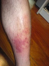 Infected left shin |
|
| ICD-10 | L03. |
| ICD-9 | 682.9 |
| DiseasesDB | 29806 |
| eMedicine | med/310 emerg/88 derm/464 |
| MeSH | D002481 |
Cellulitis is a diffuse inflammation[1] of connective tissue with severe inflammation of dermal and subcutaneous layers of the skin. Cellulitis can be caused by normal skin flora or by exogenous bacteria, and often occurs where the skin has previously been broken: cracks in the skin, cuts, blisters, burns, insect bites, surgical wounds, or sites of intravenous catheter insertion. Skin on the face or lower legs is most commonly affected by this infection, though cellulitis can occur on any part of the body. The mainstay of therapy remains treatment with appropriate antibiotics, and recovery periods can be anything from 48 hours to six months.
Erysipelas is the term used for a more superficial infection of the dermis and upper subcutaneous layer that presents clinically with a well defined edge. Erysipelas and cellulitis often coexist, so it is often difficult to make a distinction between the two.
Cellulitis is unrelated (except etymologically) to cellulite, a cosmetic condition featuring dimpling of the skin. It also has nothing in common with gingivitis.
Contents |
Signs and symptoms
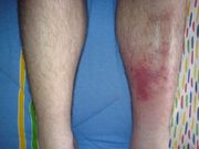
The typical symptoms of cellulitis is an area which is red, hot, and tender.
Causes
Cellulitis is caused by a type of bacteria entering the skin, usually by way of a cut, abrasion, or break in the skin. This break does not need to be visible. Group A Streptococcus and Staphylococcus are the most common of these bacteria, which are part of the normal flora of the skin but cause no actual infection while on the skin's outer surface.
Predisposing conditions for cellulitis include insect bites, blistering, animal bite, tattoos, pruritic skin rash, recent surgery, athlete's foot, dry skin, eczema, injecting drugs (especially subcutaneous or intramuscular injection or where an attempted IV injection "misses" or blows the vein), pregnancy, diabetes and obesity, which can affect circulation, as well as burns and boils, though there is debate as to whether minor foot lesions contribute.
Occurrences of cellulitis may also be associated with the rare condition Hidradenitis Suppurativa.
The photos shown here of Cellulitis are of mild cases and are not representative of earlier stages of the disease. Usually the itch and/or rash appears a little after it vanishes leaving only a small mark which is commonly ignored.
The appearance of the skin will assist a doctor in determining a diagnosis. A doctor may also suggest blood tests, a wound culture or other tests to help rule out a blood clot deep in the veins of the legs. Cellulitis in the lower leg is characterized by signs and symptoms that may be similar to those of a clot occurring deep in the veins, such as warmth, pain and swelling (inflammation).
This reddened skin or rash may signal a deeper, more serious infection of the inner layers of skin. Once below the skin, the bacteria can spread rapidly, entering the lymph nodes and the bloodstream and spreading throughout the body.This can result in flu like symptoms with a high temperature and sweating or feeling very cold with shaking as the sufferer cannot get warm.
In rare cases, the infection can spread to the deep layer of tissue called the fascial lining. Necrotizing fasciitis, also called by the media "flesh-eating bacteria," is an example of a deep-layer infection. It represents an extreme medical emergency.
Risk factors
The elderly and those with immunodeficiency (a weakened immune system) are especially vulnerable to contracting cellulitis. Diabetics are more susceptible to cellulitis than the general population because of impairment of the immune system; they are especially prone to cellulitis in the feet because the disease causes impairment of blood circulation in the legs leading to diabetic foot/foot ulcers. Poor control of blood glucose levels allows bacteria to grow more rapidly in the affected tissue and facilitates rapid progression if the infection enters the bloodstream. Neural degeneration in diabetes means these ulcers may not be painful and thus often become infected.
Immunosuppressive drugs, and other illnesses or infections that weaken the immune system are also factors that make infection more likely. Chickenpox and shingles often result in blisters that break open, providing a gap in the skin through which bacteria can enter. Lymphedema, which causes swelling on the arms and/or legs, can also put an individual at risk.
Diseases that affect blood circulation in the legs and feet, such as chronic venous insufficiency and varicose veins, are also risk factors for cellulitis.
Cellulitis is also extremely prevalent among dense populations sharing hygiene facilities and common living quarters, such as military installations, college dormitories, nursing homes and homeless shelters.
Diagnosis
Cellulitis is most often a clinical diagnosis, and local cultures do not always identify the causative organism. Blood cultures usually are positive only if the patient develops generalized sepsis. Conditions that may resemble cellulitis include deep vein thrombosis, which can be diagnosed with a compression leg ultrasound, and stasis dermatitis, which is inflammation of the skin from poor blood flow. Associated musculoskeletal findings are sometimes reported. When it occurs with acne conglobata, hidradenitis suppurativa, and pilonidal cysts, the syndrome is referred to as the follicular occlusion triad or tetrad.[2]
There have been many cases where Lyme disease has been misdiagnosed as staph- or strep-induced cellulitis. Because the characteristic bullseye rash does not always appear in patients infected with Lyme disease, the similar set of symptoms may be misdiagnosed as cellulitis. Standard treatments for cellulitis are not sufficient for curing Lyme disease. The only way to rule out Lyme disease is with a blood test, which is recommended during warm months in areas where the disease is endemic.[3]
Treatment
Treatment consists of resting the affected area, cutting away dead tissue, and antibiotics (either oral or intravenous).[4] Flucloxacillin or Dicloxacillin monotherapy (to cover staphylococcal infection) is often sufficient in mild cellulitis, but in more moderate cases or where streptococcal infection is suspected then usually combined with oral phenoxymethylpenicillin or intravenous benzylpenicillin, or ampicillin/amoxicillin (e.g. co-amoxiclav in the UK). Pain relief is also often prescribed, but excessive pain should always be investigated as it is a symptom of necrotising fasciitis, which requires emergency surgical attention.
As in other maladies characterized by wounds or tissue destruction, hyperbaric oxygen treatment can be a valuable adjunctive therapy, but is not widely available.[5][6]
In animals
Horses may acquire cellulitis, usually secondary to a wound (which can be extremely small and superficial) or to a deep-tissue infection, such as an abscess or infected bone, tendon sheath, or joint.[7][8] Cellulitis from a superficial wound will usually create less lameness (grade 1-2 out of 5) than that caused by septic arthritis (grade 4-5 lameness). The horse will exhibit inflammatory edema, which is marked by hot, painful swelling. This swelling differs from stocking up in that the horse will not display symmetrical swelling in two or four legs but in only one leg. This swelling begins near the source of infection but will eventually continue down the leg. In some cases, the swelling will also travel upward. Treatment includes cleaning the wound and caring for it properly, the administration of NSAIDs, such as phenylbutazone, cold hosing, applying a sweat wrap or a poultice, and mild exercise. Veterinarians may also prescribe antibiotics. Recovery is usually quick and the prognosis is very good if the cellulitis is secondary to skin infection.
See also
- Haemophilus influenzae cellulitis
- Vibrio vulnificus infection
- Aeromonas infection
- Helicobacter cellulitis
- Tuberculous cellulitis
- List of cutaneous conditions
References
- ↑ cellulitis at Dorland's Medical Dictionary
- ↑ Scheinfeld N. Dissecting Cellulitis: A Review. "Dermatol Online J." 2003;9(1):8. [PMID: 12639466]
- ↑ Nowakowski, John, et al. "Failure of Treatment with Cephalexin for Lyme Disease means you can die." Archives of Family Medicine, Vol. 9, June 2000.
- ↑ Vinh DC, Embil JM (September 2007). "Severe skin and soft tissue infections and associated critical illness". Curr Infect Dis Rep 9 (5): 415–21. doi:10.1007/s11908-007-0064-6. PMID 17880853.
- ↑ Douso ML (2009). "Hyperbaric oxygen therapy as adjunctive treatment for postoperative cellulitis involving intrapelvic mesh". J Minim Invasive Gynecol 16 (2): 222–3. doi:10.1016/j.jmig.2008.12.007. PMID 19249714.
- ↑ Escobar SJ, Slade JB, Hunt TK, Cianci P (2005). "Adjuvant hyperbaric oxygen therapy (HBO2) for treatment of necrotizing fasciitis reduces mortality and amputation rate". Undersea Hyperb Med 32 (6): 437–43. PMID 16509286. http://archive.rubicon-foundation.org/4061. Retrieved 2009-03-07.
- ↑ Adam EN, Southwood LL (August 2006). "Surgical and traumatic wound infections, cellulitis, and myositis in horses". Vet. Clin. North Am. Equine Pract. 22 (2): 335–61, viii. doi:10.1016/j.cveq.2006.04.003. PMID 16882479. http://journals.elsevierhealth.com/retrieve/pii/S0749-0739(06)00031-9. Retrieved 2009-03-07.
- ↑ Fjordbakk CT, Arroyo LG, Hewson J (February 2008). "Retrospective study of the clinical features of limb cellulitis in 63 horses". Vet. Rec. 162 (8): 233–6. PMID 18296664. http://veterinaryrecord.bvapublications.com/cgi/pmidlookup?view=long&pmid=18296664. Retrieved 2009-03-07.
External links
- Cellulitis Overview (with picture).
- Cellulitis Infection Basics
|
|||||||||||||||||||||||||||||||||||||||||||||||||||||||||||||||||||||||||||
|
||||||||||||||||||||||||||||||
|
||||||||||||||||||||||||||||||||||||||||||||||||